
Conditions · Stages
Varicose Vein Stages: the CEAP Classification (C0–C6)
Quick answer: Doctors grade chronic venous disease with the CEAP system. C0 means no visible signs; C1 spider or reticular veins; C2 varicose veins; C3 swelling; C4 skin changes such as darkening or hardening; C5 a healed venous ulcer; and C6 an open ulcer. Stages C3–C6 are called chronic venous insufficiency.
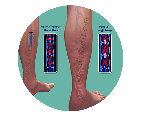
Key facts at a glance
- CEAP stands for Clinical, Etiological, Anatomical, Pathophysiological — an international system for describing venous disease.
- Clinical classes: C0 to C6, from no visible signs to an open ulcer.
- Symptom marker: “s” is added if you have symptoms (e.g. C2s), “a” if you do not.
- C2: varicose veins — bulging veins 3 mm or wider.
- C3–C6: chronic venous insufficiency.
- Progressive: untreated disease tends to move to higher classes over time.
- Staging tool: clinical examination plus duplex ultrasound.
What is the CEAP classification?
CEAP is the internationally used system for describing chronic venous disease. Its four letters stand for:
- C – Clinical: what the doctor sees on your legs (C0 to C6).
- E – Etiology: the cause — for example, primary (valves weakened on their own), secondary (after a blood clot or injury) or congenital.
- A – Anatomy: which veins are involved — superficial, deep or perforator veins.
- P – Pathophysiology: what is going wrong — reflux (backward flow), obstruction, or both.
In everyday conversation, “varicose vein stage” usually refers to the C class. The E, A and P parts come from a duplex ultrasound and help your doctor plan treatment.
What do the CEAP stages C0 to C6 mean?
C0 – No visible signs
Your legs look normal, but you may have symptoms such as heaviness or aching. Venous reflux can be found on ultrasound even at this stage.
C1 – Spider veins and reticular veins
Fine red or purple spider veins (under 1 mm) or flat blue reticular veins (1–3 mm). Often cosmetic, but sometimes a sign of reflux deeper down.
C2 – Varicose veins
Bulging, twisted veins 3 mm or wider, usually from reflux in the great or small saphenous vein or their branches. A recurrence after earlier treatment is marked C2r.
C3 – Swelling (edema)
Fluid collects around the ankles and lower leg, typically worse in the evening. See leg and ankle swelling .
C4 – Skin changes
Long-term high pressure damages the skin. C4a includes brown pigmentation and venous eczema; C4b includes lipodermatosclerosis (hard, tight skin) and atrophie blanche (white scar-like patches). See skin darkening and lipodermatosclerosis .
C5 – Healed venous ulcer
A sore has opened and healed, leaving the skin fragile and at risk of breaking down again.
C6 – Active venous ulcer
An open wound, usually near the inner ankle, that is slow to heal. See venous leg ulcers .
How does LEDAS describe the stages to patients?
LEDAS explains varicose veins to patients as a six-step progression: telangiectasia, varicose veins, edema, skin discoloration, severe discoloration and ulceration, and progressive ulcer. This maps closely to CEAP C1 through C6. The message is the same: the disease advances over time, and the earlier it is treated, the simpler treatment tends to be.
Does every patient progress through every stage?
No. Progression is not inevitable and its speed varies widely. Some people stay at C1 or C2 for many years; others develop swelling and skin changes sooner, especially with high body weight, standing work or a history of deep vein thrombosis. Varicose veins do not go away on their own, so monitoring is sensible even if you choose not to treat yet.
How does stage affect treatment?
- C0–C1: ultrasound to check for hidden reflux; sclerotherapy for spider and reticular veins; lifestyle measures and compression if symptomatic.
- C2–C3: close the refluxing trunk veins with EVLT and treat branches with duplex-guided sclerotherapy.
- C4–C6: treat reflux to lower pressure on the skin. Where skin is damaged, LEDAS recommends ultrasound-guided sclerotherapy first, as surgery becomes harder. Compression supports healing.
Treatment cost depends on stage. At LEDAS, the average is about 5,500,000 KRW per leg, with an exact quote after ultrasound.
CEAP clinical classes at a glance
| Class | What you see | Common symptoms | Typical approach at LEDAS |
|---|---|---|---|
| C0 | No visible veins | Heaviness, aching possible | Ultrasound if symptomatic |
| C1 | Spider or reticular veins | Often none; sometimes burning | Ultrasound check, sclerotherapy |
| C2 | Bulging varicose veins ≥ 3 mm | Aching, heaviness, cramps | EVLT + duplex-guided sclerotherapy |
| C3 | Ankle and leg swelling | Tight shoes, evening swelling | Treat reflux; compression |
| C4 | Darkening, eczema, hard skin | Itching, tenderness | Treat reflux promptly |
| C5 | Healed ulcer scar | Fragile skin | Treat reflux to prevent recurrence |
| C6 | Open ulcer | Pain, discharge | Ultrasound-guided sclerotherapy first; wound care |
Varicose vein stages – frequently asked questions
At what CEAP stage should varicose veins be treated?
There is no single cut-off. Treatment is usually recommended from C2 when there are symptoms or confirmed reflux on ultrasound, and strongly advised from C3 onward to prevent skin damage. Spider veins at C1 can be treated for comfort or appearance after an ultrasound check.
Can varicose veins go back to an earlier stage?
Swelling (C3) often improves after the leaking veins are treated, and some skin darkening may fade slowly. Hardened skin and ulcer scars usually remain, though they are protected from getting worse. This is why earlier treatment generally gives a better outcome.
What does “C2s” or “C2r” mean on my report?
“s” means you have symptoms such as aching or heaviness, and “a” means you have none. “r” marks recurrent varicose veins after previous treatment. Your report may also list E, A and P codes describing cause, veins involved and type of flow problem.
How quickly do varicose veins progress?
It varies from person to person. Some stay stable for years; others progress faster, especially with obesity, standing work or a previous blood clot. Regular review, ideally with ultrasound, is the best way to know. Read are varicose veins dangerous?
About this page
Clinical information:
LEDAS Varicose Vein Clinic, led by
Dr. Kim Byoung Joon , thoracic and cardiovascular surgeon, founding member of the Korean Society for Phlebology.
Medical reviewer:
Dr. Kim Byoung Joon ·
Review status:
Pending ·
Last updated:
October 2026
This page is general information and not a diagnosis. Treatment is decided only after an in-person examination. Read our
editorial policy .
Related: Varicose veins guide · Chronic venous insufficiency · Symptoms · Venous leg ulcers · Treatment planning
Find out your stage with a duplex ultrasound
English consultation available · +82-70-4895-5086 · kbjledaspm@gmail.com
