
Conditions · Skin changes
Skin Discoloration from Varicose Veins and Lipodermatosclerosis
Quick answer: Brown or reddish-brown skin around the ankles and lower calves is a sign that long-standing vein reflux is damaging the skin. Iron from leaked red blood cells stains the tissue, and over time the skin can harden and tighten (lipodermatosclerosis). It marks an advanced stage of vein disease, and treating the underlying reflux helps protect the skin from ulcers.
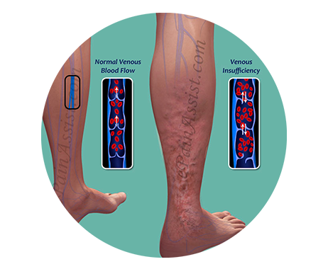
Key facts at a glance
- Hemosiderin staining: brown discoloration from iron left behind when red blood cells leak out of high-pressure veins.
- Lipodermatosclerosis: inflammation and scarring of the skin and fat under it, making the lower leg hard, tight and sometimes narrowed like an inverted bottle.
- Venous eczema: itchy, dry, flaky or weeping skin over the ankles, often the first skin change.
- Stage: skin discoloration is stage 4 of the 6-stage progression LEDAS describes; ulcers follow at stages 5–6.
- Typical location: inner ankle and lower calf.
- What helps: treating the reflux, compression, and good skin care.
Why do varicose veins darken the skin?
In healthy legs, valves inside the veins keep blood moving toward the heart. When they fail, blood flows backward and collects near the ankle. This creates long-term high pressure in the smallest vessels, called venous hypertension .
Under this pressure, fluid and red blood cells slowly leak into the skin. When the red cells break down, they leave behind an iron-containing pigment called hemosiderin . It stains the skin brown, a bit like a bruise that never fully fades. The leaked material also triggers inflammation, which is why the area may itch, feel warm or look red.
Skin discoloration sits in the middle of the disease progression described in the LEDAS varicose vein stages : after visible veins and swelling, and before ulcers. It is part of chronic venous insufficiency .
What is lipodermatosclerosis?
Lipodermatosclerosis (LDS) means inflammation and hardening (sclerosis) of the skin (derma) and the fat beneath it (lipo). It develops in two phases:
Acute phase
The lower leg becomes red, warm, tender and painful. It is sometimes mistaken for cellulitis (a skin infection), but there is usually no fever and antibiotics do not help.
Chronic phase
Over months, inflammation gives way to scarring. The skin turns brown and feels hard and tight, as if it is stuck to the tissue underneath. The lower leg may narrow just above the ankle while the calf stays full, an "inverted champagne bottle" shape. Skin in this state is fragile and slow to heal.
What other skin changes come with vein disease?
- Venous (stasis) eczema: itchy, dry, scaly or weeping patches near the ankle. LEDAS lists frequent itching near the ankle and dry, itchy calf skin among varicose vein symptoms.
- Atrophie blanche: small, smooth, white scar-like areas, often with tiny red dots, which can be painful.
- Corona phlebectatica: a fan of fine blue or purple veins around the inner ankle or foot.
- Easy bruising and skin that does not lighten over time.
Is skin discoloration dangerous?
The color itself is not dangerous, but it is a warning sign. It shows the skin has been under strain for a long time and is closer to breaking down into a venous leg ulcer . LEDAS notes that once skin necrosis or ulcers develop, surgery becomes more difficult, which is why acting at the discoloration stage makes sense.
See a doctor promptly if the skin becomes hot, red and spreading with fever, if a wound opens and does not heal within two weeks, or if one leg suddenly swells and hurts, which may be a DVT .
How does LEDAS treat vein-related skin changes?
- Map the reflux. A standing duplex ultrasound on Philips EPIQ Elite equipment finds the leaking veins, including perforator veins that often feed the skin of the ankle.
- Treat gently through healthier skin. Because damaged skin is fragile, LEDAS favors non-surgical approaches. Ultrasound-guided foam sclerotherapy reaches the refluxing veins through a fine needle, and EVLT may close a larger saphenous vein from a puncture away from the damaged area. All under local anesthesia, with no incision.
- Support the skin. Medical compression stockings , daily moisturizing, and treating eczema flare-ups with your doctor.
- Follow up. Ultrasound every one to two years to find new reflux early.
Will the brown color fade?
Once the pressure is relieved, inflammation settles and the skin often feels softer. Hemosiderin staining fades slowly and may not disappear completely, especially if it has been present for years. The main goal of treatment is to protect the skin from further damage and ulcers.
Vein-related skin changes at a glance
| Change | What you see or feel | What it means |
|---|---|---|
| Venous eczema | Itchy, dry, flaky or weeping skin near ankle | Early skin reaction to vein pressure |
| Hemosiderin staining | Brown patches that do not fade | Long-standing leakage of red blood cells |
| Acute lipodermatosclerosis | Red, warm, tender lower leg | Active inflammation of skin and fat |
| Chronic lipodermatosclerosis | Hard, tight, narrowed lower leg | Scarring; high risk of ulcer |
| Atrophie blanche | White, smooth, scar-like spots | Fragile skin; ulcers heal slowly here |
Skin darkening and lipodermatosclerosis – frequently asked questions
Can creams fix brown skin from varicose veins?
Moisturizers and prescribed steroid creams can calm itching and eczema, but they do not remove the cause. The staining comes from blood leaking under pressure. Treating the refluxing veins is what lowers that pressure; creams are a helpful addition, not a replacement.
Is lipodermatosclerosis the same as cellulitis?
No, although they can look similar. Cellulitis is a bacterial infection, often with fever, that needs antibiotics. Acute lipodermatosclerosis is inflammation from vein disease, usually on both sides of the lower leg, without fever. If you are unsure, see a doctor promptly.
Can varicose veins be treated when the skin is already hard?
Yes, in most cases. Ultrasound-guided foam sclerotherapy can close the veins beneath hardened skin through a fine needle, without cutting it. LEDAS generally prefers this non-surgical route when the skin is damaged, adding laser where suitable after the ultrasound.
Why is the discoloration always near my inner ankle?
The inner ankle is the lowest point where the great saphenous vein and several perforator veins meet, so pressure there is highest when you stand. That is why skin changes and ulcers usually start in this "gaiter" area.
About this page
Clinical information:
LEDAS Varicose Vein Clinic, led by
Dr. Kim Byoung Joon , thoracic and cardiovascular surgeon, founding member of the Korean Society for Phlebology.
Medical reviewer:
Dr. Kim Byoung Joon ·
Review status:
Pending ·
Last updated:
October 2026
This page is general information and not a diagnosis. Treatment is decided only after an in-person examination. Read our
editorial policy .
Related: Venous leg ulcers · Chronic venous insufficiency · Leg and ankle swelling · Complications of untreated varicose veins · Foam sclerotherapy
Protect your skin before it breaks down
English consultation available · +82-70-4895-5086 · kbjledaspm@gmail.com
