
Conditions · Reticular Veins
Reticular Veins: What They Are and How They're Treated
Quick answer: Reticular veins are flat blue or green veins, 1–3 mm wide, that sit just under the skin, often behind the knee, on the outer thigh or around the ankle. They are larger than spider veins but smaller than varicose veins, and often feed spider-vein clusters. LEDAS treats them with sclerotherapy after an ultrasound check.
Key facts at a glance
- Size: 1–3 mm wide — between spider veins and varicose veins.
- Look: flat, blue-green, net-like (“reticular” means net-like).
- CEAP class: C1.
- Common sites: behind the knee, outer thigh, inner calf, ankle.
- Role: often act as “feeder veins” for spider veins.
- Treatment at LEDAS: sclerotherapy (SCT), after ultrasound to check for deeper reflux.
What are reticular veins?
Reticular veins are small, widened veins in the deeper layer of the skin. They are 1–3 mm wide, look blue or blue-green, and usually lie flat rather than bulging. The word “reticular” means “net-like”, which describes how they often form a branching network. You may also hear them called “feeder veins” or “blue veins”.
In the CEAP classification , reticular veins sit in class C1 together with spider veins . When a vein reaches 3 mm or more and becomes raised and twisted, it is classed as a varicose vein (C2).
Why do reticular veins matter?
Reticular veins are often the hidden supply line for spider veins. A cluster of fine red or purple spider veins usually has one or more reticular veins nearby feeding blood into it. If only the spider veins are treated, they often come back, because the feeder is still carrying pressure into the area.
Reticular veins can also be connected to reflux in larger veins further up or deeper in the leg. That is why LEDAS recommends a duplex ultrasound before treating them, even when they look minor.
What causes reticular veins?
- Inherited weakness in vein walls and valves
- Female hormones, pregnancy and menopause
- Standing or sitting for long periods
- Ageing and loss of vein elasticity
- Excess weight and an inactive lifestyle
Thin, fair skin also makes reticular veins more visible. Learn more on what causes varicose veins .
Do reticular veins cause symptoms?
Often they do not. Some people feel mild aching, burning or itching over them, especially after standing or in the heat. If you have heaviness, ankle swelling or night cramps as well, ask for an ultrasound, as these suggest reflux in larger veins.
How are reticular veins treated?
Step 1: Ultrasound check
Dr. Kim Byoung Joon scans the leg, standing, to see whether the reticular veins are linked to a refluxing trunk or perforator vein.
Step 2: Treat any deeper source
If larger veins are leaking, they are treated first, usually with EVLT for trunk veins or duplex-guided sclerotherapy for hidden veins.
Step 3: Sclerotherapy of the reticular veins
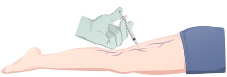
A fine needle injects a sclerosing agent — as liquid or foam — into the reticular vein. The lining becomes irritated, the vein closes and the body absorbs it over the following weeks. Treating the feeder vein first often makes nearby spider veins fade too. See sclerotherapy for spider and reticular veins and foam vs liquid sclerotherapy .
Step 4: Compression and walking
Wearing medical compression stockings and walking lightly for 2–4 weeks helps the vein close and reduces bruising.
What is recovery like?
Sclerotherapy takes a few minutes and you can return to normal activities straight away. Treated veins may look darker or feel firm for a few weeks before fading, which is a normal part of healing. Mild bruising and temporary brown marks can occur. Avoid hot baths, saunas and intense exercise for a short time as advised.
Reticular vein treatment pathway at LEDAS
| Step | What happens | Why |
|---|---|---|
| 1. Duplex ultrasound | Standing scan of superficial, perforator and deep veins | Finds hidden reflux feeding the area |
| 2. Treat deeper reflux (if any) | EVLT or duplex-guided sclerotherapy | Removes the pressure source |
| 3. Sclerotherapy | Liquid or foam injected into reticular veins | Closes the feeder veins |
| 4. Compression & walking | Stockings and light walking for 2–4 weeks | Helps veins close and fade |
| 5. Review | Check results; treat remaining spider veins | Completes the cosmetic and medical result |
Reticular veins – frequently asked questions
Are reticular veins the same as varicose veins?
No. Reticular veins are smaller (1–3 mm), flat and blue-green, and are classed as CEAP C1. Varicose veins are 3 mm or wider, raised and twisted, and classed as C2. Reticular veins can, however, be linked to the same underlying reflux, so an ultrasound check is sensible.
Are reticular veins dangerous?
Reticular veins themselves are not dangerous. They rarely cause complications. Their importance is that they can feed spider veins and sometimes signal reflux in larger veins. Treatment is usually for comfort or appearance, after ruling out deeper disease.
Why are my blue veins more visible lately?
Blue veins can become more visible with weight loss, ageing skin, pregnancy, hormonal changes or warm weather. New or enlarging veins, especially with aching or swelling, are worth checking with ultrasound. Read when to see a vein doctor.
How many sessions do reticular veins need?
Many people see good improvement after one or two sessions, but it depends on how many veins there are and whether spider veins are also treated. Sessions are spaced weeks apart. See how many sclerotherapy sessions do I need?
About this page
Clinical information:
LEDAS Varicose Vein Clinic, led by
Dr. Kim Byoung Joon , thoracic and cardiovascular surgeon, founding member of the Korean Society for Phlebology.
Medical reviewer:
Dr. Kim Byoung Joon ·
Review status:
Pending ·
Last updated:
October 2026
This page is general information and not a diagnosis. Treatment is decided only after an in-person examination. Read our
editorial policy .
Related: Spider veins · Sclerotherapy · Foam vs liquid sclerotherapy · Varicose veins guide · CEAP stages
Treat blue reticular veins at the source
English consultation available · +82-70-4895-5086 · kbjledaspm@gmail.com
